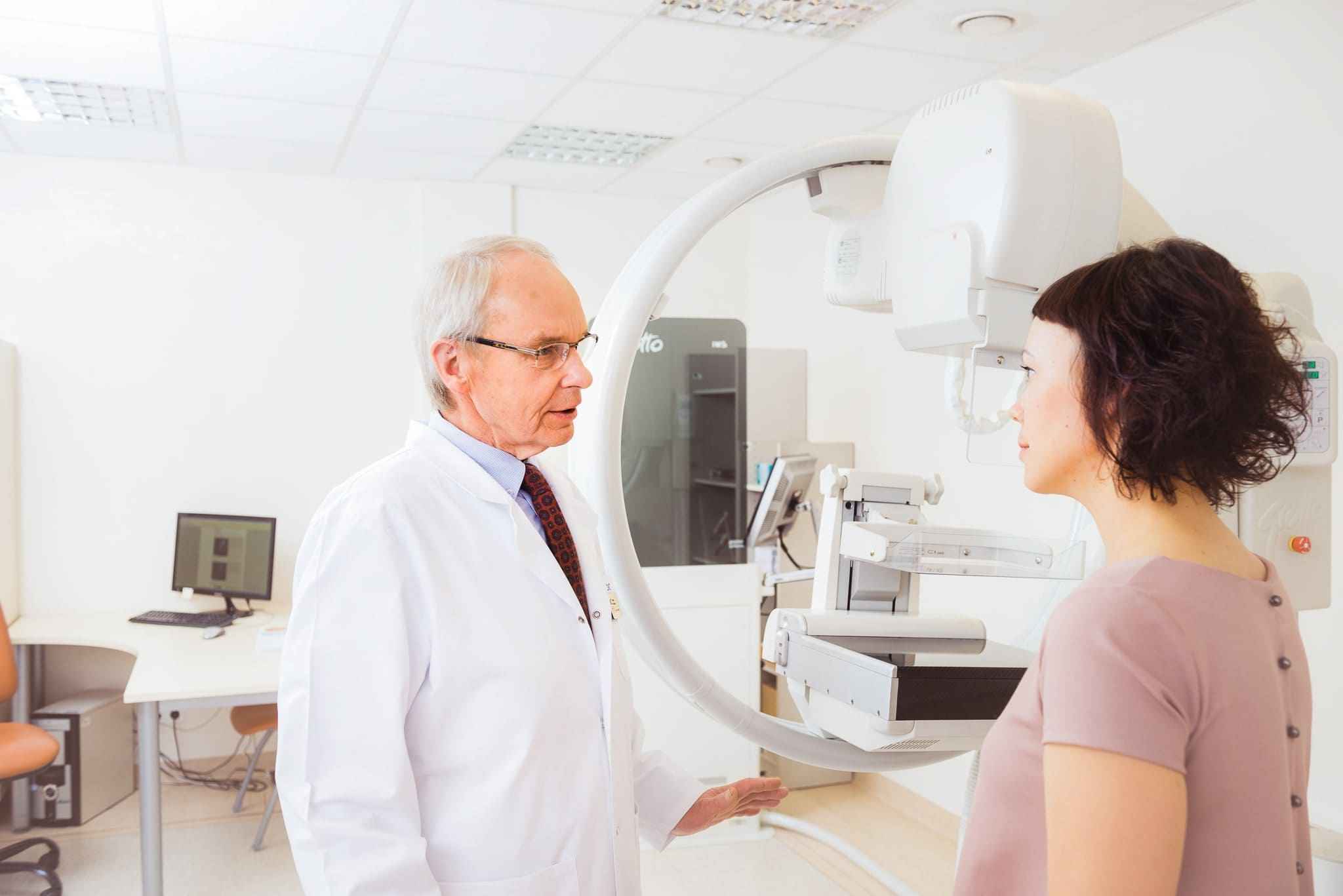
Mammography
Sorry, we are not providing mammography services temporarily.
Breast cancer is one of the most common oncological diseases among women both in Lithuania and worldwide. It is important to be aware that breast cancer can be cured entirely, if diagnosed in its early stages. After a check-up, you can feel calm all year.
A mammography examination helps in diagnosing malignant and benign lesions of the breast tissue.
Who should have a mammography examination?
- women over the age of 40 once every two years
- women whose mother or grandmothers had breast cancer (examinations should start 10 years before the age when breast cancer was diagnosed in the family)
For reliable protection against breast cancer, mammography is combined with a breast ultrasound examination.
All women over the age of 20 should have a check-up once per year even if they have no complaints and regularly examine their breasts by palpation.
Keep in mind that quite often the disease starts quietly, so if you have a check-up you will protect yourself.
Examinations are performed by radiologists.
The examination lasts approximately 15 to 30 minutes, and the results are given immediately afterwards.
Price of mammography examination
What factors affect the price?
The prices indicated below apply to citizens of the Republic of Lithuania and the European Union.
If you are coming from another country please check the price by telephoning or sending an email.
Mammography unit Giotto Image 3DL
At the Radiology Centre, which is one of the divisions of the Medical Diagnostic and Treatment Centre, mammography examinations are carried using a Giotto Image 3DL (Italy) digital unit. The circle shape is very comfortable for a seated patient, and during the examination she can relax her pectoral muscles even more, resulting up to 2 cm more breast tissue being visualised compared to other mammography units. More accurate examinations can help save women’s lives. A good example is the former first lady of the United States, Barbara Bush, who was diagnosed with a breast tumour especially close to the thorax using a Giotto mammography unit. More about the manufacturer of the Giotto Image 3DL mammography unit.


- Minimum X-ray irradiation; 2 cm more breast tissue visualised compared to examinations by other mammography units.
- Examination is performed by qualified specialists with extensive experience.
- Modern, safe and reliable equipment by world-leading manufacturers.
- We can schedule your visit in such a way that the test is carried out, a consultation by the necessary specialist is provided and treatment is prescribed all on the same day.
Useful to know
What are the advantages of having an examination carried out by our mammography unit?
- the examination can be performed in a position usual for the patient or face to face, which allows the operator to better see the patient at the time of positioning
- due to its unique form, 2 cm more breast tissue is visualised compared to examinations using other mammography units
The mammography procedure is painless, because the Giotto Image 3DL unit has a special sensitive breast compression system installed that is controlled by computer:
- speedy movement to the breast
- automatic stop before the breast compression
- compression is regulated by the breast’s density and the machine adapts to the breast being examined
- slow and even release of compression after the examination
This mammography unit enables the examination of both standing and sitting patients, i.e. those who are immobile.
When is mammography contraindicated?
Mammography is an X-ray examination and consequently is not performed during pregnancy. Furthermore, it is not suitable for very young females whose mammary gland tissue is dense and does not conduct the X rays well. For the same reason, it is best to perform the mammography in the first week after menstruation.


